Description
Validation Data
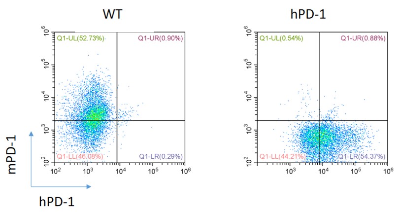
Figure 1. Expression of PD-1 in the activated spleen lymphocytes of homozygous humanized PD-1 BALB/c KI mice is detected by FACS.
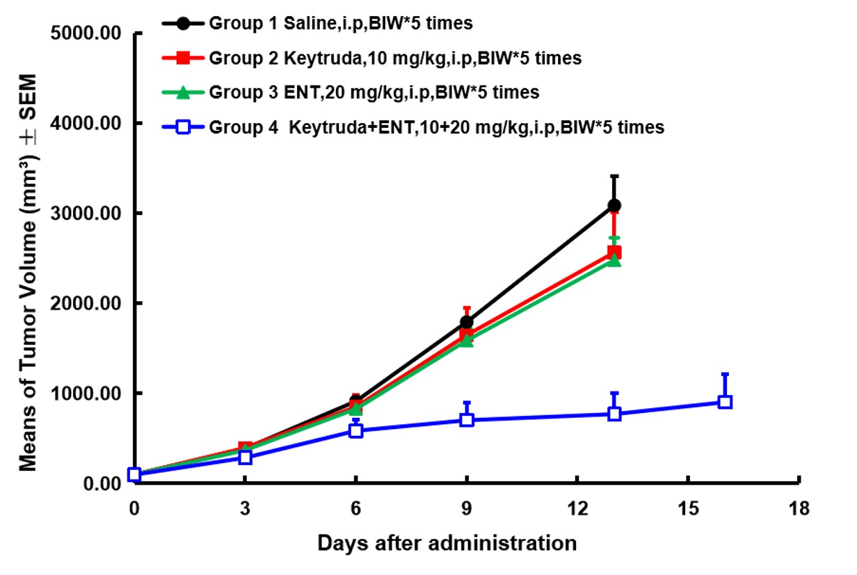
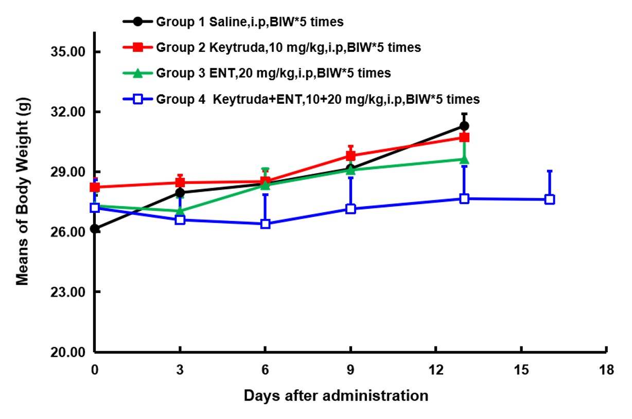
Figure 2. In vivo validation of homozygous BALB/c-hPD1 mice. The homozygous BALB/c-hPD1 mice were inoculated with CT26 cells, and randomly assigned to different groups (n=7) when the tumor grew to a volume of 100 mm3. A combinatorial treatment of anti-hPD1 antibody Keytruda and entinostat (ENT; a class I HDAC inhibitor) demonstrated a noticeable efficacy improvement compared to the same dose of single agent (A) without affecting the animal body weight (B).
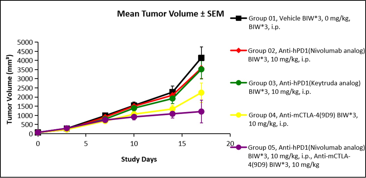
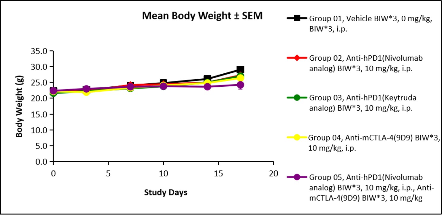
Figure 3. In vivo validation of homozygous BALB/c-hPD1 mice. The homozygous BALB/c-hPD1 mice were inoculated with CT26 cells, and randomly assigned to different groups (n=6) when the tumor grew to a volume of 65 mm3. A combinatorial treatment of anti-hPD1 antibody Opdivo and anti-mCTLA antibody demonstrated a noticeable efficacy improvement compared to the same dose of single agent (A) without affecting the animal body weight (B). (Data in partnership with collaborators)


Reviews
There are no reviews yet.