
Generation Strategy

Figure 1. Generation strategy of humanized PD-1 mice. On the C57BL/6J genetic background, the protein coding sequences for human PDCD1 gene were inserted into the ATG position of the mouse Pdcd1 gene, so that the expression of endogenous Pdcd1 in the mouse was replaced by the expression of full length PDCD1 protein of human.
Data from Flow Cytometry (FACS) Analysis
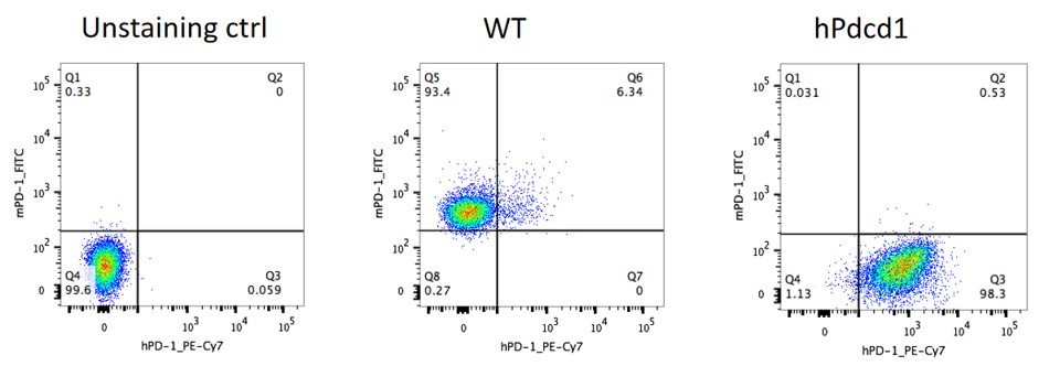
Figure 2. Expression of PD-1 in the activated spleen lymphocytes of humanized PD-1 Homozygous mice is detected by FACS.
In Vivo Validation in a MC38 Tumor-bearing Model of the Humanized PD-1 Mice
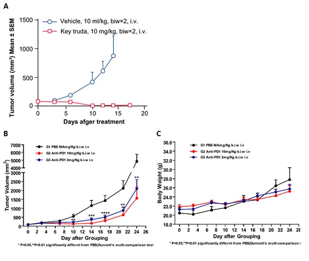
Figure 3. In vivo anti-tumor effect of an anti-human PD-1 antibody in a humanized mouse model of PD-1. Anti-human-PD-1 drugs significantly inhibited the growth of MC38 tumors in PD-1 mice, demonstrating that the humanized PD-1 mice can be used to assess the anti-human PD-1 antibody.
A. Mean volume ± SEM of tumor tissues ((Data in partnership with collaborators) In vivo validation of anti-tumor efficacy in a MC38 tumor-bearing model of humanized PD-1 mice. Homozygous humanized PD-1 mice were inoculated with MC38 colon cancer cells. After the tumors grew to 100 mm3, the animals were randomly assigned into a control group and a treatment group (n=8). The drug was given twice a week for a total of 4 administrations. The results showed that Keytruda, a drug targeting human PD-1, exerted a very significant anti-tumor effect (p<0.001), demonstrating that the humanized PD-1 mice are a good in vivo model for validating the efficacy of antibodies targeting human PD-1.
B. Mean volume ± SEM of tumor tissues. C. Mean body weight ± SEM of mice ((Data in partnership with collaborators)
In vivo dose validation of anti-tumor efficacy in a MC38 tumor-bearing model of humanized PD-1 mice. Homozygous humanized PD-1 mice were inoculated with MC38 colon cancer cells. After the tumors grew to about 90 mm3, the animals were randomly assigned into a control group and a treatment group (n=9). The results showed that the antibodies targeting human PD-1 showed a very significant antitumor effect (p<0.001), and such antitumor effect is dose-dependent.
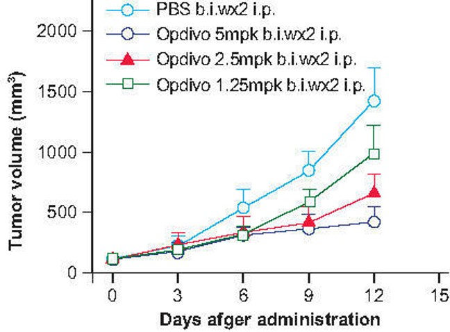
Figure 4. In vivo dose validation in a MC38 tumor-bearing model of humanized PD-1 mice. Homozygous humanized PD-1 mice were inoculated with MC38 colon cancer cells. After the tumors grew to 100 mm3, the animals were randomly assigned into a control group and a treatment group (n=8). The drug was given twice a week for a total of 4 administrations.


Reviews
There are no reviews yet.