
Validation Data
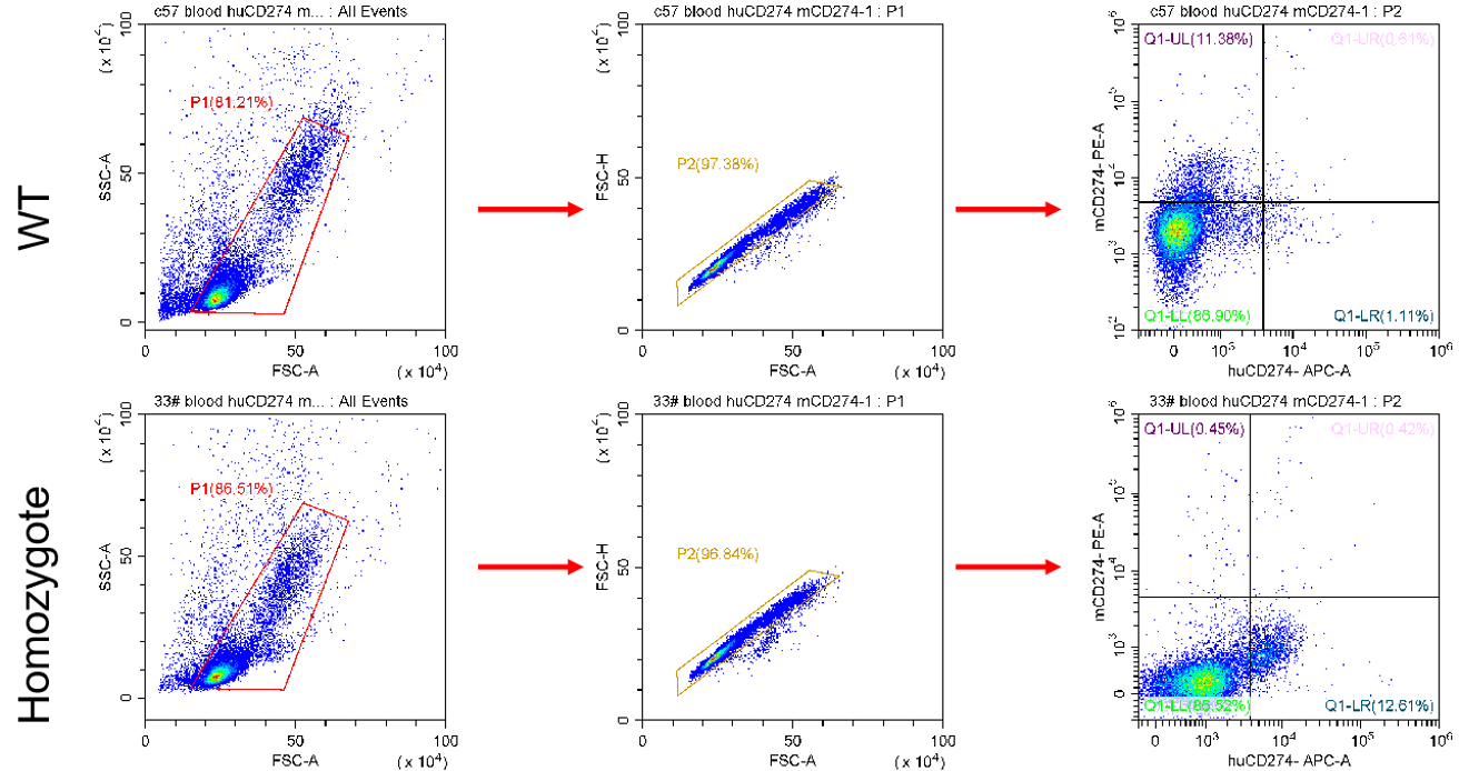
Figure 1. The expression of human PD-L1 in the peripheral blood cells derived from homozygous humanized mice were confirmed by flow cytometry analysis.
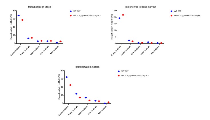
Figure 2. Immunotype in blood , spleen and bone marrow in hPD-L1 mice.
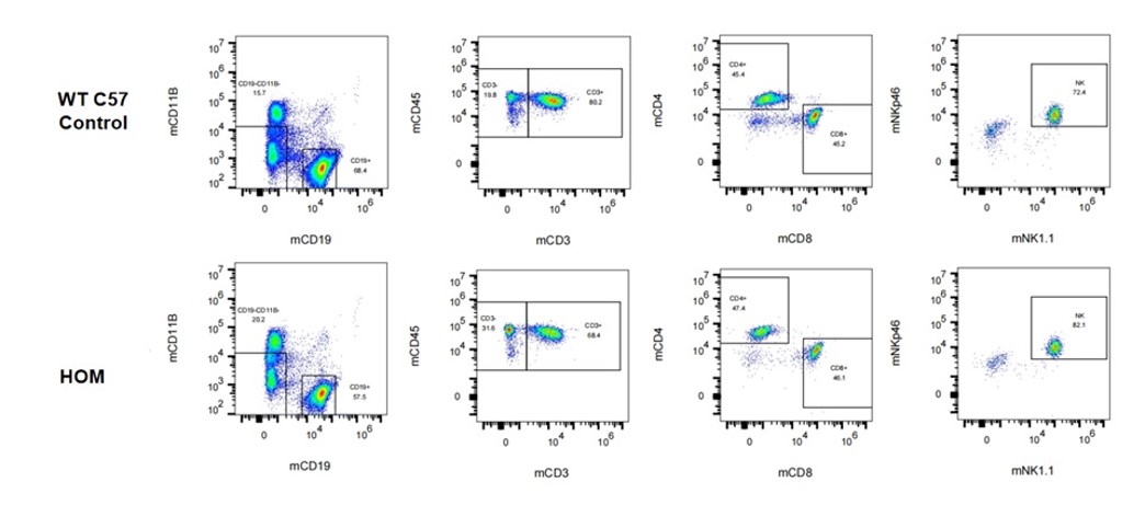
Figure 3. Immunotype in blood in hPD-L1 mice.
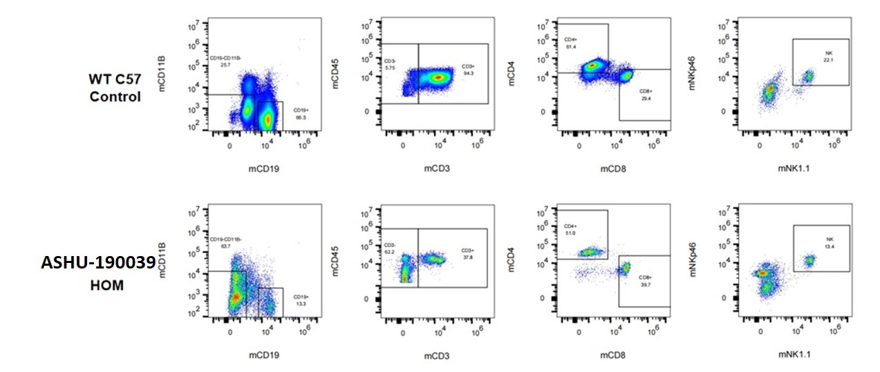
Figure 4. Immunotype in spleen in hPD-L1 mice.
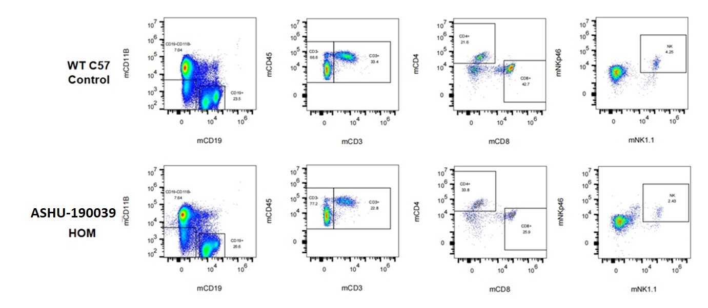
Figure 5. Immunotype in bone marrow in hPD-L1 mice.
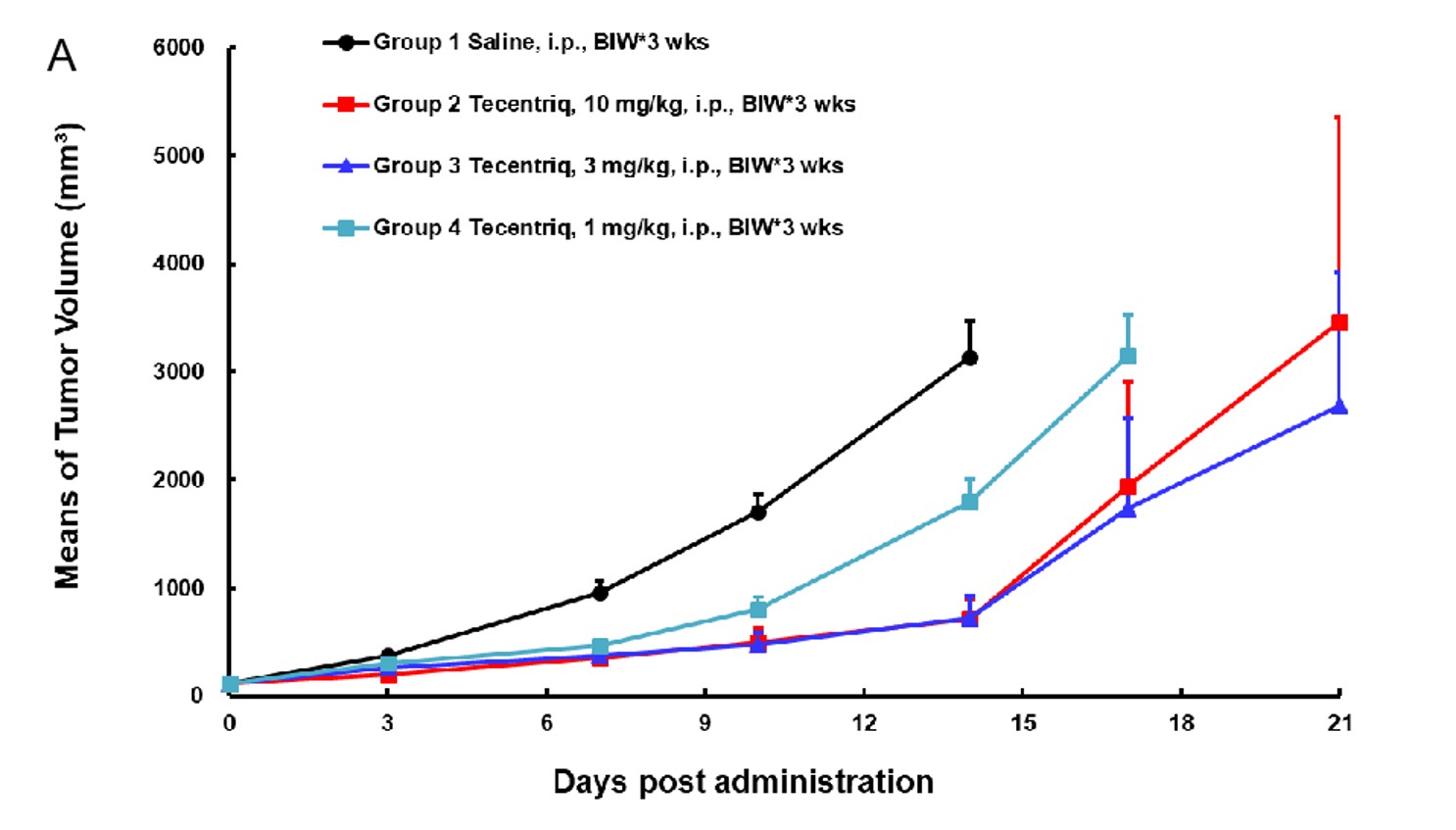
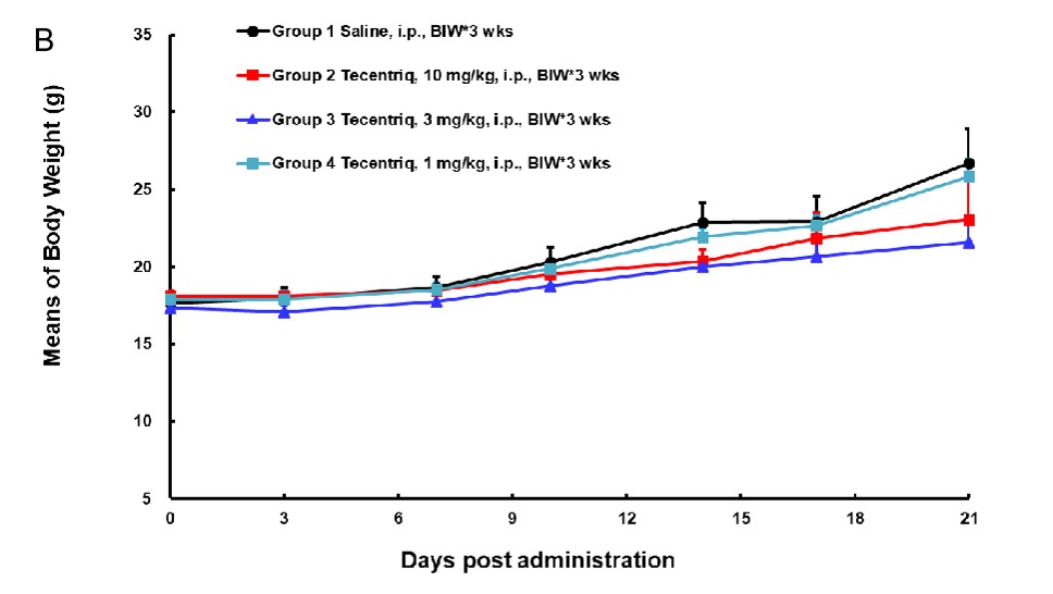
Figure 6. In vivo anti-tumor effect of an anti-human PD-L1 antibody in the humanized PD-L1 mouse model. (Tecentriq is a monoclonal antibody specific for human PD-L1)
(A) PD-L1 humanized MC38 cells were subcutaneously implanted into humanized PD-L1 mouse model. Treatment of hPD-L1 antibody inhibited PD-L1 humanized MC38 tumor growth, demonstrating that the humanized PD-L1 mice are a good in vivo model for validating the efficacy of antibodies targeting human PD-L1.
(B) Mean body weight of mice. All quantitative data are represented as mean ± SEM.


Reviews
There are no reviews yet.